

Cell Fragment Formation
Cell Fragment Formation, Migration, and Force Exertion on Extracellular Mimicking Fiber Nanonets
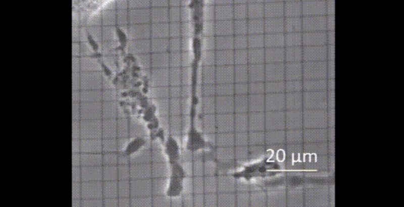
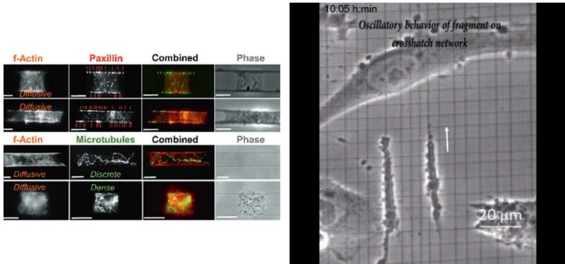
Cell fragments devoid of the nucleus play an essential role in intercellular communication. Mostly studied on flat 2D substrates, their origins and behavior in native fibrous environments remain unknown. Here, cytoplasmic fragments’ spontaneous formation and behavior in suspended extracellular matrices mimicking fiber architectures (parallel, crosshatch, and hexagonal) are described. After cleaving from the parent cell body, the fragments of diverse shapes on fibers migrate faster compared to 2D. Furthermore, while fragments in 2D are mostly circular, a higher number of rectangular and blob‐like shapes are formed on fibers, and, interestingly, each shape is capable of forming protrusive structures. Absent in 2D, fibers’ fragments display oscillatory migratory behavior with dramatic shape changes, sometimes remarkably sustained over long durations (>20 h). Immunostaining reveals paxillin distribution along fragment body‐fiber length, while Forster Resonance Energy Transfer imaging of vinculin reveals mechanical loading of fragment adhesions comparable to whole cell adhesions. Using nanonet force microscopy, the forces exerted by fragments are estimated, and peculiarly small area fragments can exert forces similar to larger fragments in a Rho‐associated kinase dependent manner. Overall, fragment dynamics on 2D substrates are insufficient to describe the mechanosensitivity of fragments to fibers, and the architecture of fiber networks can generate entirely new behaviors.
Abinash Padhi